Summary
Milk vesicles share their size range with casein micelles, and MISEV2023 names casein as the greatest difficulty in separating vesicles from milk, especially milk from cows and other ruminants. A light-scatter particle count cannot tell a casein micelle from a vesicle of the same size, so a count from milk means little without the separation steps behind it. In one bovine milk study, two isolation methods gave about the same diameter, 120.5 and 124.3 nm, but only one of the two fractions was rich in vesicle markers.
Why are milk vesicles hard to separate?
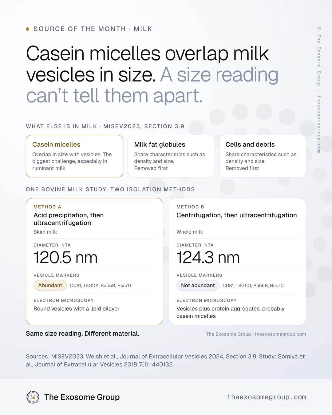
Besides vesicles, milk holds cells, milk fat globules, casein micelles and a great deal of dissolved protein. MISEV2023, the reporting guideline from the International Society for Extracellular Vesicles, gives milk its own section, 3.9, and sets out the order of difficulty. Milk fat globules and cell debris share vesicle characteristics such as density and size and should be removed first, for example by centrifugation. It describes casein micelles, which overlap in size with vesicles, as the biggest difficulty, especially for milk of ruminant species.
Casein is also abundant: in cow’s milk it makes up about 80 percent of the total protein, as Rahman and colleagues note (PLoS One, 2019), so a preparation from bovine milk starts with a large casein load to remove.
| Component in milk | What it shares with vesicles | How it is usually handled |
|---|---|---|
| Cells and cell debris | Characteristics such as density and size | Removed early, for example by centrifugation |
| Milk fat globules | Characteristics such as density and size | Removed early, for example by centrifugation |
| Casein micelles | Overlap in size with vesicles | Precipitated with acid at pH 4.6, aggregated with enzymes, or dissociated with EDTA or sodium citrate |
What does casein do to a particle count?
Casein micelles are counted along with the vesicles. Nanoparticle tracking analysis in its usual light-scatter mode follows particles moving in a liquid and reports how many it detects in a size window, without information about what each particle is made of. MISEV2023 warns that nanoparticle tracking of complex biofluids “should be interpreted with caution due to counting of co-isolates,” and recommends that methods unable to tell vesicles from other particles report a “particle” concentration rather than an “EV” concentration.
The authors of the study described below consider casein micelles similar to milk vesicles in their colloidal properties, citing a diameter of about 100 nm and a density of about 1.08 g/mL for the micelles (Somiya et al., 2018). A particle of that size and density travels with vesicles in a centrifuge and appears beside them in a size reading, and What a particle count measures covers the general form of this gap.
Can a size reading tell a clean fraction from a casein-laden one?
A size reading did not distinguish them in the bovine milk study by Somiya and colleagues in the Journal of Extracellular Vesicles (2018). They prepared vesicles in two ways, from skim milk treated with acetic acid to precipitate casein and then ultracentrifuged, and from whole milk taken through centrifugation and ultracentrifugation without the acid step. Both preparations measured about 120 nm in diameter by nanoparticle tracking, at 120.5 and 124.3 nm in their Table 1.
| What was compared | Acid precipitation, then ultracentrifugation | Centrifugation, then ultracentrifugation |
|---|---|---|
| Starting milk | Skim milk | Whole milk |
| Diameter by nanoparticle tracking | 120.5 nm | 124.3 nm |
| Vesicle marker proteins (CD81, Rab5B, TSG101, Hsc70) | Abundant | Not abundant |
| Electron microscopy | Round vesicles with a lipid bilayer | Vesicles plus protein aggregates, probably casein micelles |
| Proteins identified | 605 | 180 |
The two fractions gave similar size readings while containing different material, so the size figure missed the difference between them that the marker panel and the micrographs detected.
A count from milk cannot be read on its own, because it depends on what the separation left behind, and only the method section says what that was.
Does removing casein change the vesicles?
Removing casein can change the vesicles. MISEV2023 lists acidification to pH 4.6, enzymatic aggregation, and calcium sequestration with EDTA or sodium citrate as ways to deal with casein. It says that acidification and EDTA are used most often and that there is currently no preferred method. It also notes that single-step approaches give low purity, and that ultracentrifugation, density gradients and size-exclusion chromatography may be combined for higher purity.
Each of these steps has a cost. Rahman and colleagues compared ultracentrifugation alone with hydrochloric acid and acetic acid treatment of bovine milk (PLoS One, 2019). The acid treatments gave significantly higher particle concentrations but partly degraded the surface markers CD9 and CD81, while CD63, MFG-E8 and the internal proteins HSP70 and Alix showed no change. Some acid-treated vesicles also had rough surfaces under electron microscopy.
The step that removes the most casein may also change which markers can be read afterward, and that possibility is a reason to report the step in full.
Other factors that change a milk count
- MISEV2023 notes that colloidal properties and acceptable storage times may differ for raw, homogenized, pasteurized, ultra-high temperature-treated, and dried or powdered milk, so processing can change the count.
- The guideline also notes that the effects of storage length and temperature on milk have yet to be fully determined, and it recommends keeping milk at body temperature for short-term storage before processing.
- Vesicles have been separated from the milk of at least 16 species, chiefly human and bovine, and MISEV2023 singles out ruminant milk for its casein content.
Does this apply to colostrum?
We expect the same questions to apply to colostrum, but the studies cited here used milk and the milk section of MISEV2023 does not discuss colostrum separately, so we apply them to colostrum by inference. For any colostrum ingredient, its own method section settles the question.
How common is milk on exosome product pages?
Milk is rare on these pages so far. In our Product Index of 168 products from 73 brands, recorded from their product pages on 4 November 2026, 2 products are counted under milk or colostrum, because milk is the first source their pages name. A third product lists milk exosomes in its ingredient list after a microbial source and is counted as microbial, since each product is counted once, by the first source its page names. The figure is low partly by design, because products that mention milk exosomes only in their ingredient lists were not added as rows in this first pass. Our shorter note, Milk vesicles and casein, covers the core of the separation problem.
Limits of this note
- We did not measure any milk-derived product, so this note cannot show whether one contains vesicles, or how many.
- MISEV2023 names no best method for removing casein, and the choice depends on what will be measured afterward.
- We do not assess whether any product works.
What to request from a milk ingredient supplier
For a milk- or colostrum-derived ingredient, ask the supplier how the vesicles were separated before asking for the count, and specifically:
- How cells and milk fat were removed, with the centrifugation settings
- How casein was removed or disrupted: acid, enzyme, EDTA or citrate, and at which step
- Which separation steps followed, and whether more than one was combined
- Whether casein and fat were tracked through the process, and how
- Which vesicle markers were tested, and whether the casein step could have affected them
- The milk’s species and processing state: raw, pasteurized, ultra-high temperature-treated or powdered
A count that arrives with those answers can be interpreted. Without them, a count is a size reading of an unknown mixture.
The Index records what each page says without evaluating the product that the page sells.
If you are working with a milk-derived topical ingredient and would like to discuss the separation step, you can email us at hello@theexosomegroup.com.
Last reviewed . This note gives general scientific information about measurement and is not legal or regulatory advice or an assessment of any product.
Sources
- Welsh JA et al. Minimal information for studies of extracellular vesicles (MISEV2023). J Extracell Vesicles. 2024;13(2):e12404.
- Somiya M, Yoshioka Y, Ochiya T. Biocompatibility of highly purified bovine milk-derived extracellular vesicles. J Extracell Vesicles. 2018;7(1):1440132. PMID 29511463.
- Rahman MM et al. Acidification effects on isolation of extracellular vesicles from bovine milk. PLoS One. 2019;14(9):e0222613.
- The Exosome Group Product Index: 168 products from 73 brands, recorded from their product pages on 4 November 2026. Each product’s page uses exosome or extracellular vesicle wording. Published as distributions only; no brand, product or page is named.