Summary
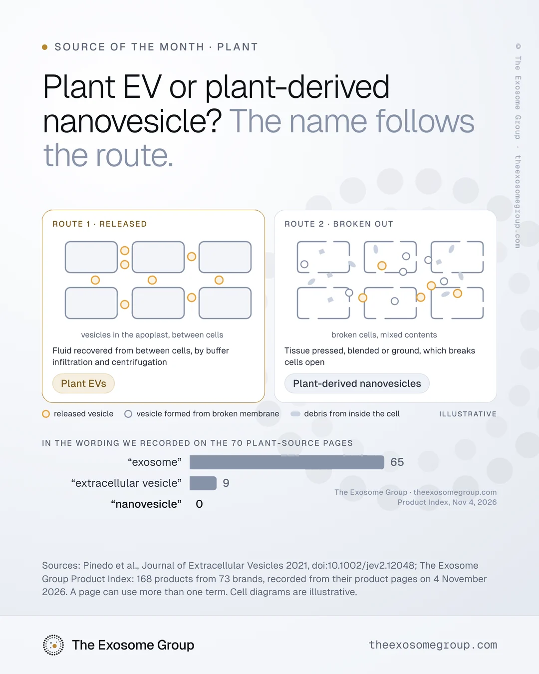
“Plant EVs” are vesicles released by plant cells into the space between them, the apoplast, whereas “plant-derived nanovesicles” are vesicle fractions recovered by pressing, blending or grinding plant tissue, where natural release cannot be shown. Pinedo and colleagues proposed the two names in 2021 because the routes give different material: breaking tissue adds artificial vesicles from broken membranes and vesicles from inside the cell. A particle count or a size reading cannot tell the two apart, so the first question for a plant ingredient is the route.
What is a plant EV?
Plant cells release vesicles into the apoplast, the fluid-filled space outside the cell membranes. Researchers recover that fluid without breaking the tissue, conventionally by infiltrating seeds or leaves with a buffer and then centrifuging the fluid back out. The buffer has to be optimized to keep tissue damage and intracellular contamination low (Pinedo et al., 2021).
In a 2021 call for rigor and standardization in plant vesicle research, published in the Journal of Extracellular Vesicles, Pinedo and colleagues recommended the generic term “plant EVs” for these vesicles rather than “exosomes.” That recommendation holds at least until further characterization allows classification by where in the cell the vesicles formed.
What is a plant-derived nanovesicle?
Most published plant vesicle work, the same authors noted, used methods likely to rupture tissue, such as extracting juice by pressing fruit or grinding it in a mixer or blender. Gentle pressing might bring only mild contamination from broken cells, but blending, they wrote, will recover EVs together with artificial particles formed from re-mixed broken membranes and native intracellular vesicles released when cells are broken open.
For material made this way, they proposed “plant-derived nanovesicles,” or PDNVs, for all vesicle fractions obtained from plant tissues when destructive processes are used and natural release into the extracellular space cannot be established.
| Plant EVs | Plant-derived nanovesicles | |
|---|---|---|
| How the material is obtained | Recovered from the apoplastic fluid, for example by buffer infiltration and centrifugation of leaves | Pressing, blending or grinding tissue, often with juice extraction |
| What comes with it | Extracellular material, with as little intracellular content as the method allows | Released vesicles, artificial vesicles from broken membranes, and vesicles from inside the cell |
| When the name applies | Natural release into the extracellular space can be established | Destructive processes were used and natural release cannot be established |
| Terms to avoid | “Exosomes,” until the vesicles’ origin in the cell is characterized | “Exosomes,” “plant EVs” and “exosome-like” terms |
Are plant EVs and plant-derived nanovesicles different materials?
They differ in the comparison that Pinedo and colleagues describe. Liu and colleagues isolated vesicles from Arabidopsis with the same isolation procedure from two starting materials, disrupted leaf and apoplastic fluid (Liu et al., Nanomedicine, 2020). As Pinedo and colleagues summarize it, the two shared some characteristics but differed in size, density distribution and protein content. The study identified 1,438 distinct proteins in the leaf nanovesicles and 787 in the apoplastic EVs, and only a small portion of the leaf nanovesicle proteins were also found in the EVs.
Some protein families, such as HSP70, GAPDH, glutathione S-transferase and annexins, turn up in both kinds of preparation, which is expected because a disrupted-tissue preparation contains whatever EVs were present along with everything else.
Can a particle count or size reading tell them apart?
A count or size reading cannot separate them, because both routes give membrane vesicles in overlapping size ranges. Pinedo and colleagues add a further problem: mixtures from disrupted tissue are often passed through 0.2 or 0.4 µm filters, which positively select, or even produce, particles in the 30 to 200 nm range expected for native EVs. A size distribution that looks like an EV preparation can be partly the work of the filter.
Markers do not yet settle the question either. Candidate plant EV markers include PEN1, PEN3 and TET8, but none is agreed as universal, and the authors noted that most antibodies against the plant candidates were not commercially available when they wrote. Animal-cell markers are the wrong first test for plant exosomes covers the marker question.
What does MISEV2023 say about plant material?
MISEV2023, the reporting guideline from the International Society for Extracellular Vesicles, has no section for plants; its source sections cover sources for which the society has or recently had a task force, and it invites proposals for new ones. Its general definitions still point the same way as Pinedo’s naming, defining an EV as a particle released from cells, delimited by a lipid bilayer, that cannot replicate on its own. MISEV2023 notes that particles from a cell that has been extruded have not strictly been released, and suggests names such as “EV mimetics” for EV-like particles produced through direct disruption of cells. Whatever the name, it asks that the name indicate the production process without implying a particular biogenesis pathway.
It also discourages “exosome” for any vesicle population whose origin in the cell’s endosomal system has not been shown, and for plant material made by blending or pressing, “exosome” describes neither the route nor the origin.
What do plant exosome pages call their material?
Most of them call it “exosomes.” In our Product Index of 168 products from 73 brands, recorded from their product pages on 4 November 2026, 70 pages claim a plant source, and the wording we recorded from those pages uses the following terms:
| Term in the recorded wording | Plant-source pages |
|---|---|
| “exosome” or “exosomes” | 65 |
| “extracellular vesicle” | 9 |
| “nanovesicle” | 0 |
Many pages pair a plant name with words such as “extracted,” “isolated” or “derived from.” Those words name a starting material, but they do not say whether the vesicles were released by living cells or recovered by breaking tissue.
Why the route is relevant to a buyer
The route determines what the material is. Neither name ranks the material, but the two carry different contents, so evidence gathered on one does not transfer to the other, and a document should say which one it describes. A supplier who uses both terms for the same ingredient has a question to answer before any count is read.
Limits of this note
- This note does not show whether any plant ingredient contains vesicles, or which kind, since we measured no product.
- The route determines the name and the questions to ask, and it does not decide whether one ingredient is better than another.
- We do not assess whether any product works.
Questions for a plant ingredient supplier
- The plant, by species and tissue
- The route, whether apoplastic fluid, pressing, blending, grinding or another process, described step by step
- Whether any filtration step was used, and the pore size
- Which name the supplier uses for the material, and why
- Which markers were tested, with the reagent named
- Electron microscopy showing membrane-bound vesicles, and an estimate of what else was recovered
The Index records what each page says and does not evaluate the product that the page describes.
If your supplier’s documents use both terms, you can email us at hello@theexosomegroup.com.
Last reviewed . This note gives general scientific information about measurement and is not legal or regulatory advice or an assessment of any product.
Sources
- Pinedo M et al. A call for rigor and standardization in plant extracellular vesicle research. J Extracell Vesicles. 2021;10(6):e12048.
- Liu Y et al. Characterization of and isolation methods for plant leaf nanovesicles and small extracellular vesicles. Nanomedicine. 2020;29:102271. PMID 32702466. Protein comparison cited as summarized in Pinedo et al., 2021.
- Welsh JA et al. Minimal information for studies of extracellular vesicles (MISEV2023). J Extracell Vesicles. 2024;13(2):e12404.
- The Exosome Group Product Index: 168 products from 73 brands, recorded from their product pages on 4 November 2026. Each product’s page uses exosome or extracellular vesicle wording. Published as distributions only; no brand, product or page is named.